


While in training at the University of Iowa's ophthalmology department, I was fortunate enough to have Dr. Stephen R. Russell as one of my mentors. Dr. Russell, a vitreoretinal surgeon and researcher, had himself trained at Iowa, and developed a love for retinal drawings.
In the past, ophthalmologists would often spend 30 minutes or more drawing a picture of what they saw during the patient's eye exam. The retina, the thin layer that lines the inside of the back of your eye, is the most expressive part of the eye, and even the exact same disease can look different from one patient to the next.
Since the advent of improved ophthalmic photographic techniques -- and the increase in the number of patients needing to be seen each day -- ophthalmologists don't take nearly as much time to draw their findings anymore. Lamenting this, Dr. Russell put together a beautiful book containing many exquisite drawings done by dozens of different ophthalmologists at Iowa.
Looking through this book for the first time was an emotional experience. Taking care of people's eyes means so much to me, and I could literally feel the love that these doctors -- all of whom had walked the same halls I was then walking, and many of whom had gone on to become giants in our profession -- had for their patients and their craft as they made these meticulous, beautiful drawings. I decided to put a little more effort into my drawings as time allowed.
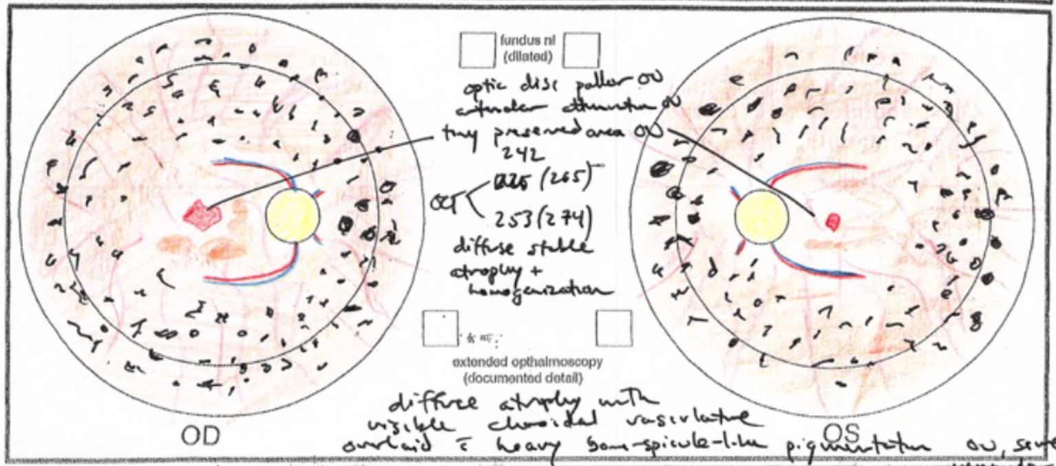
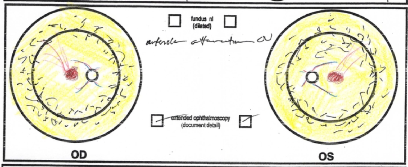
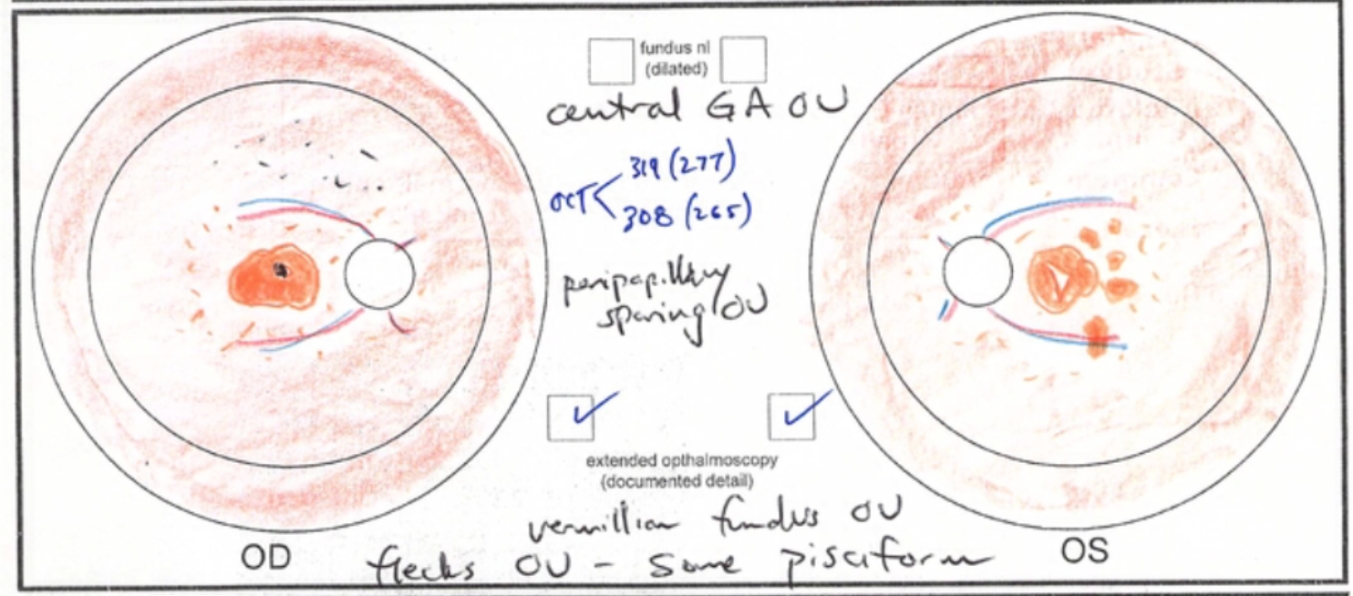
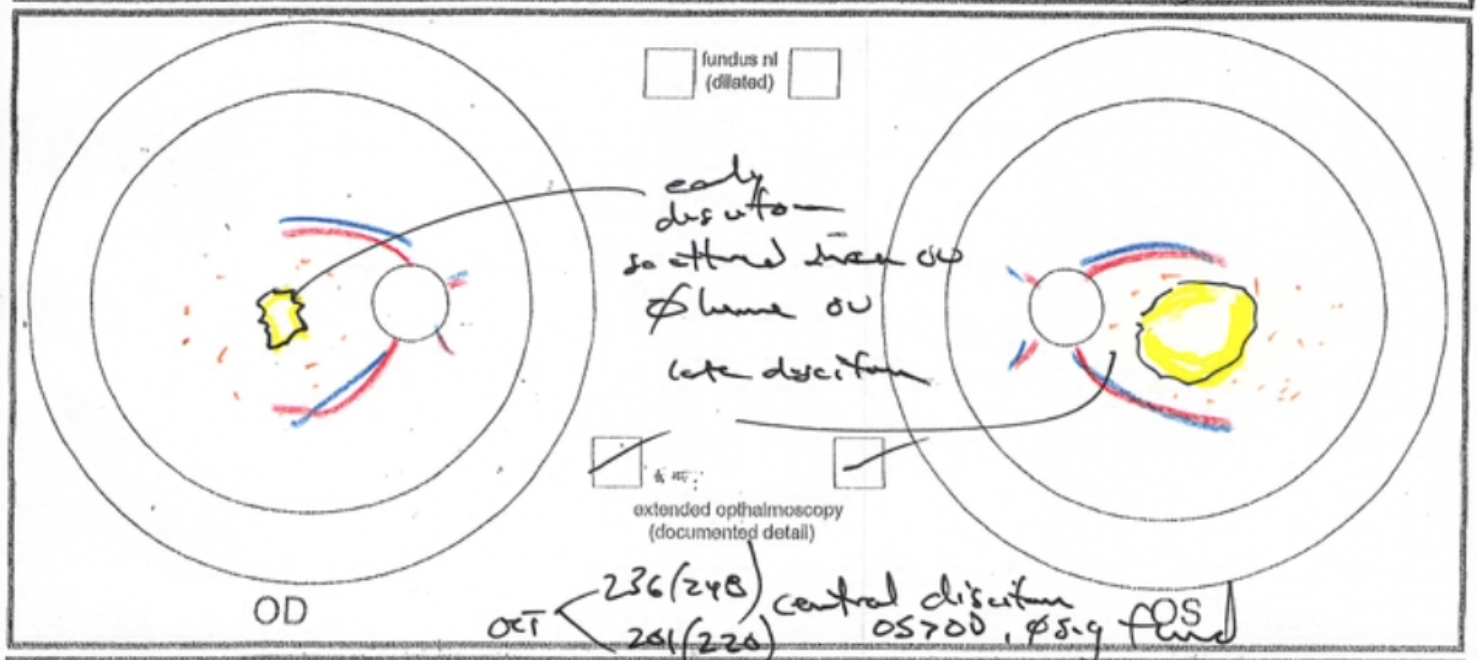
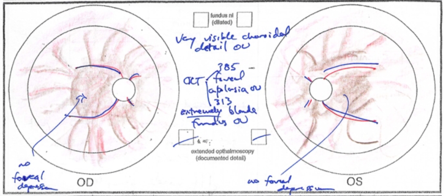
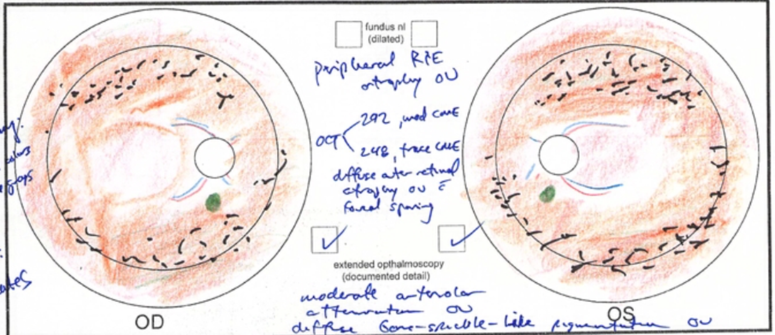
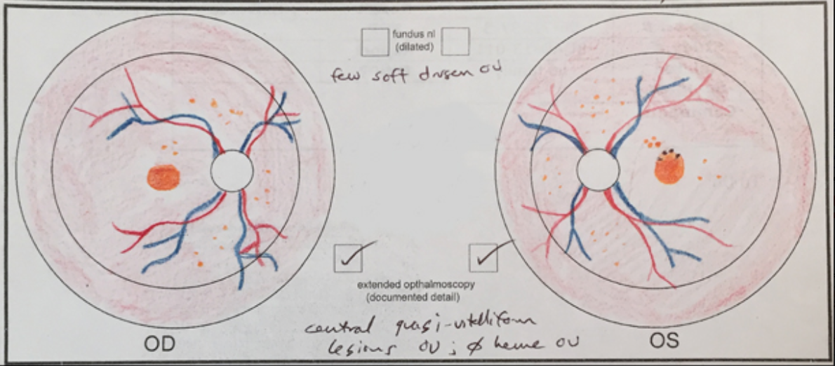
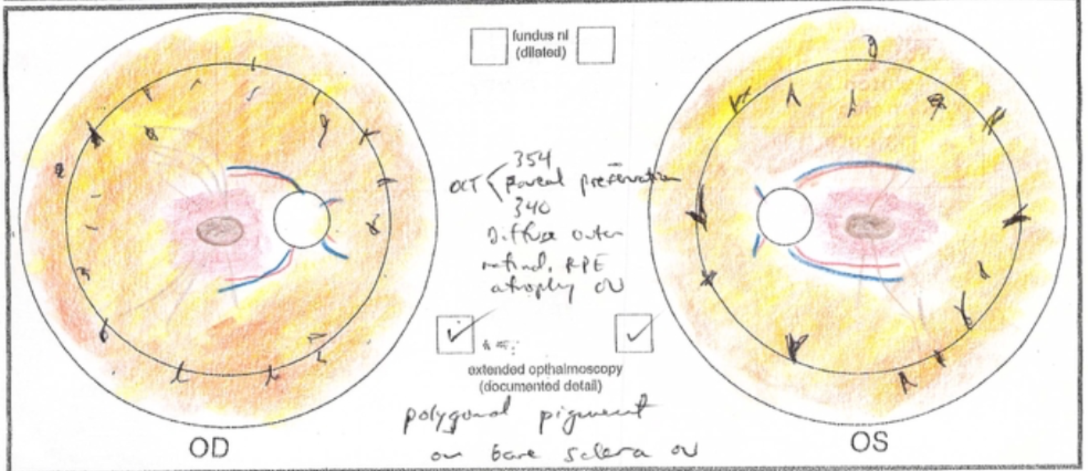
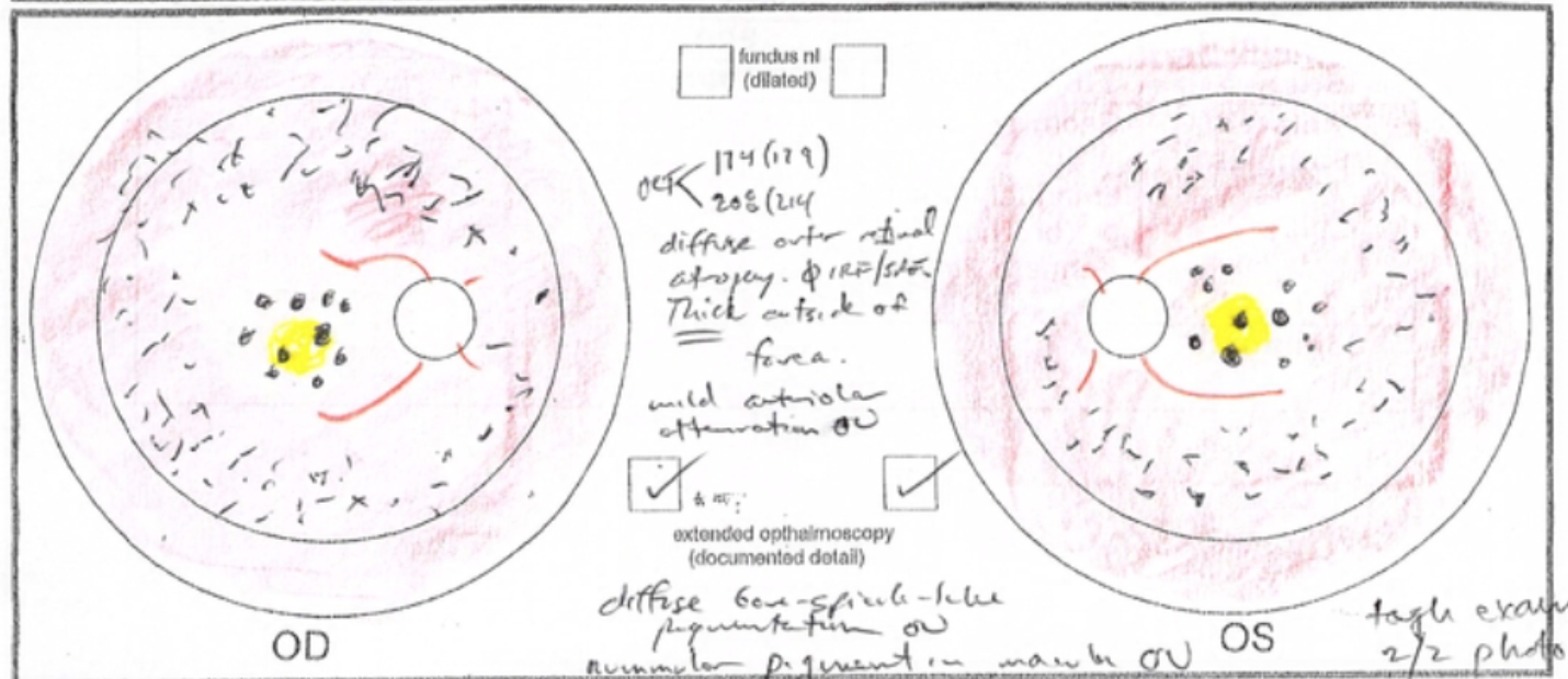
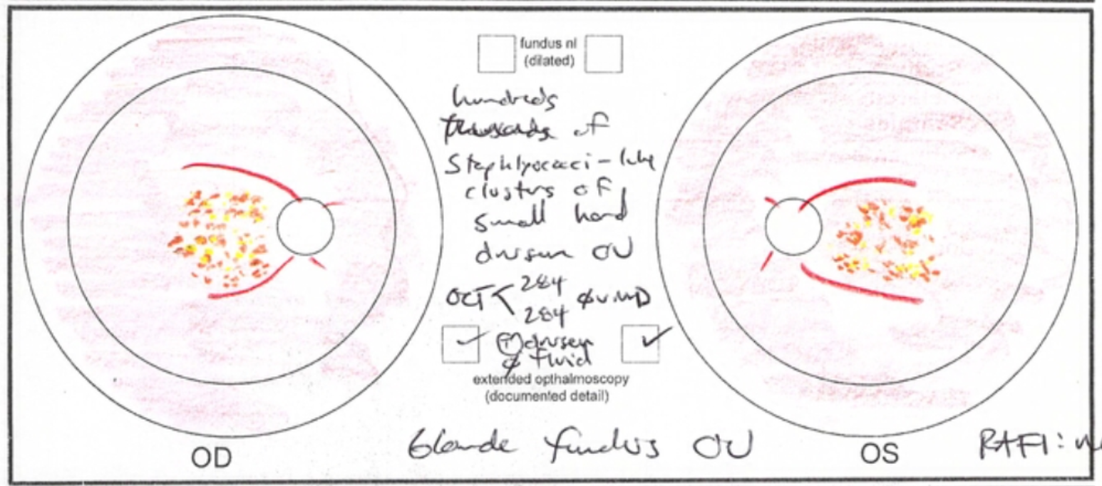
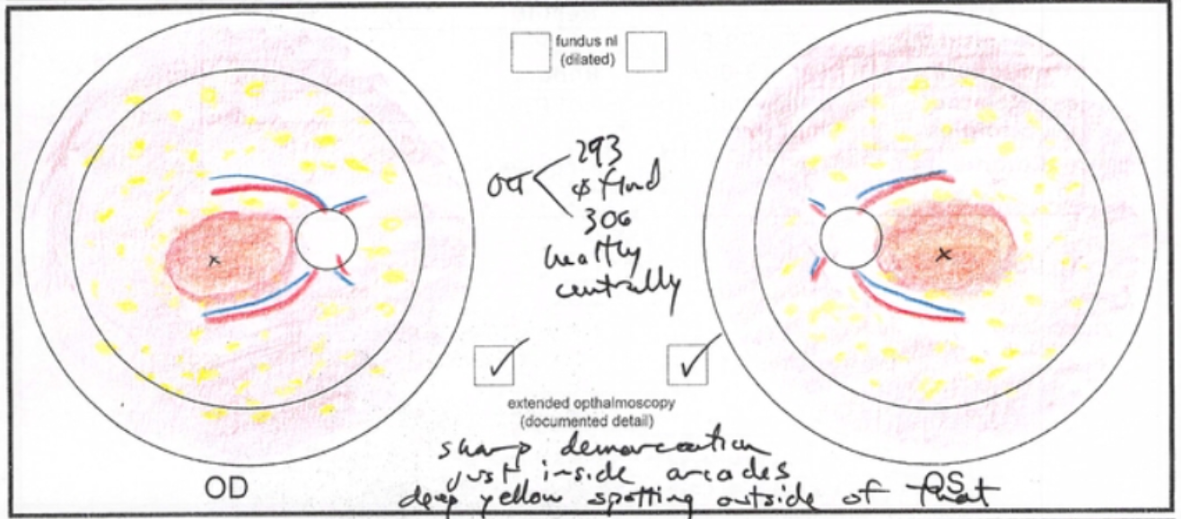
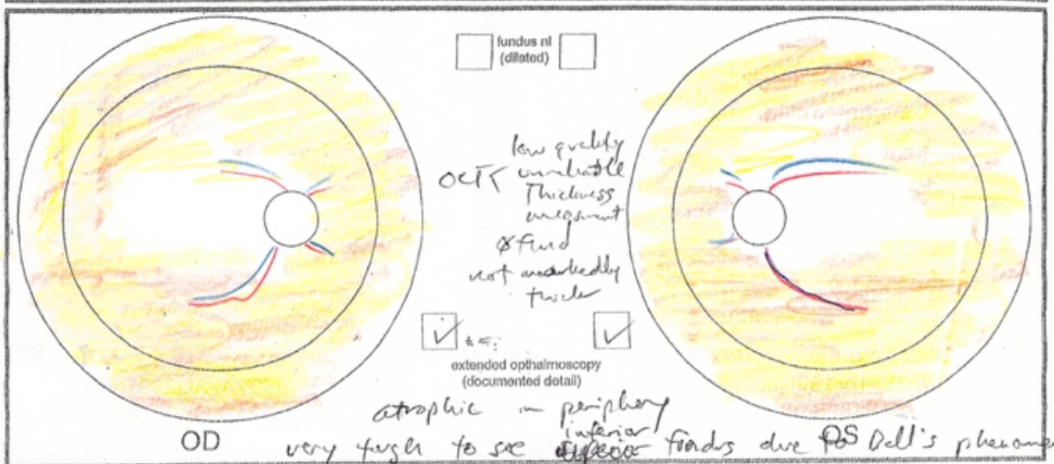
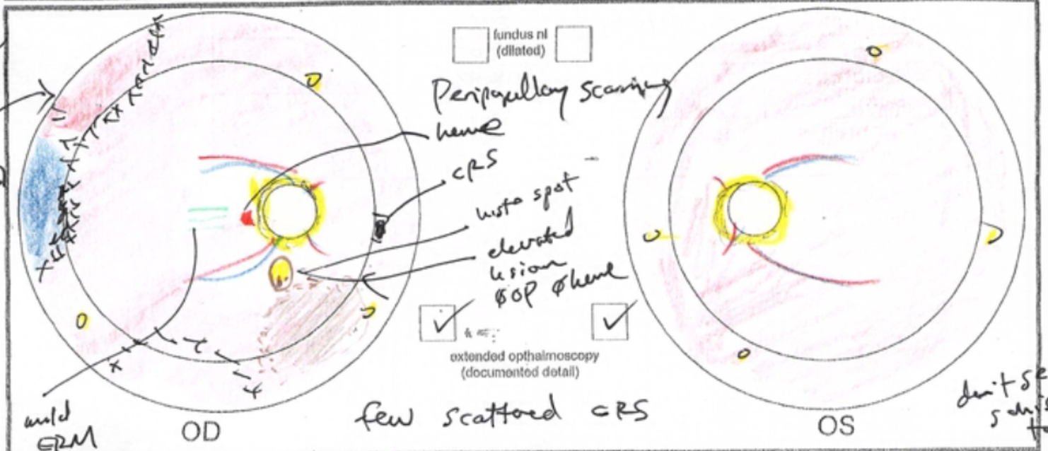
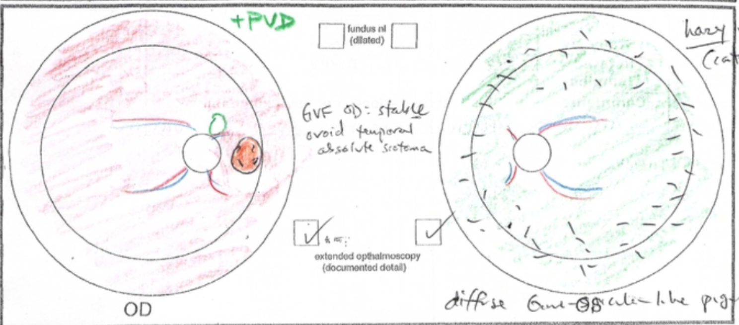
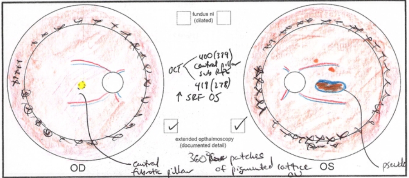
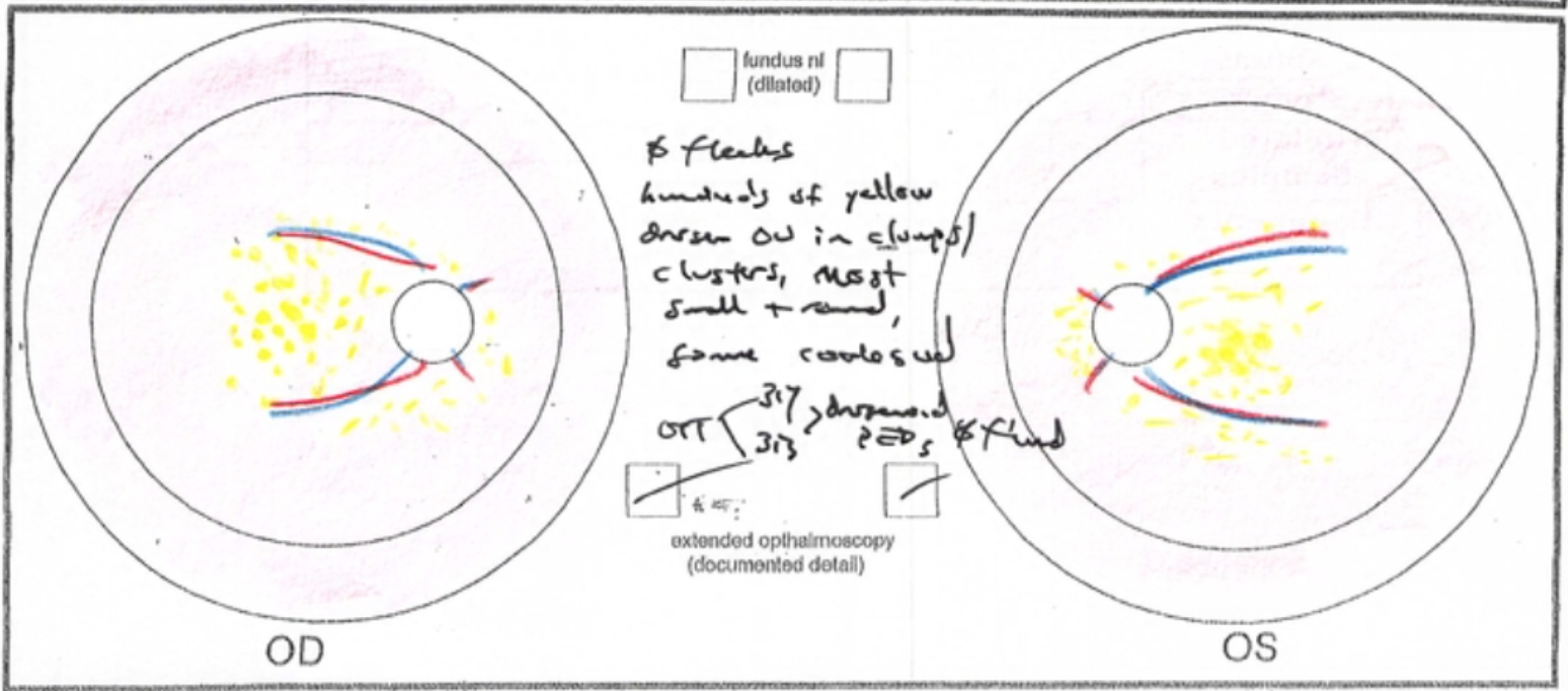
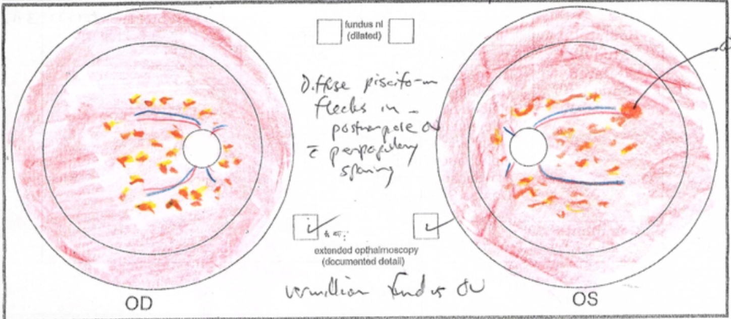
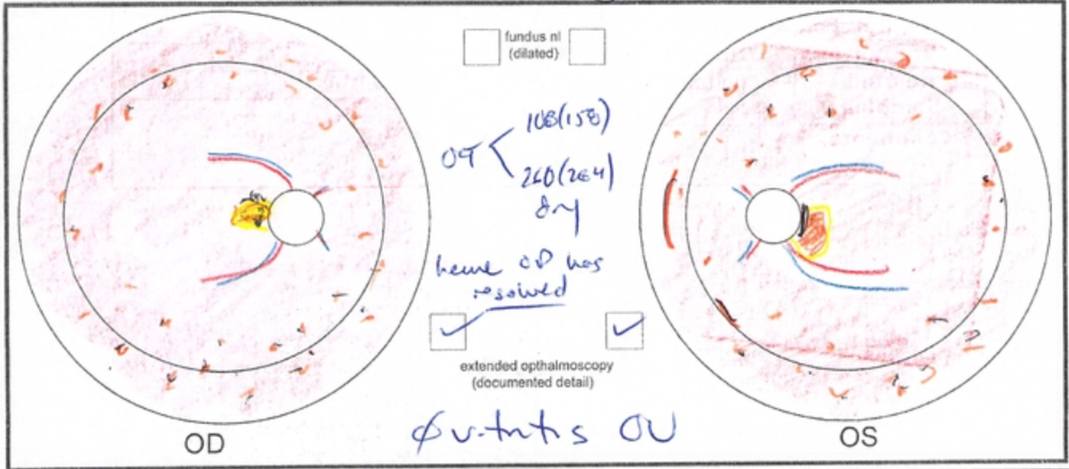
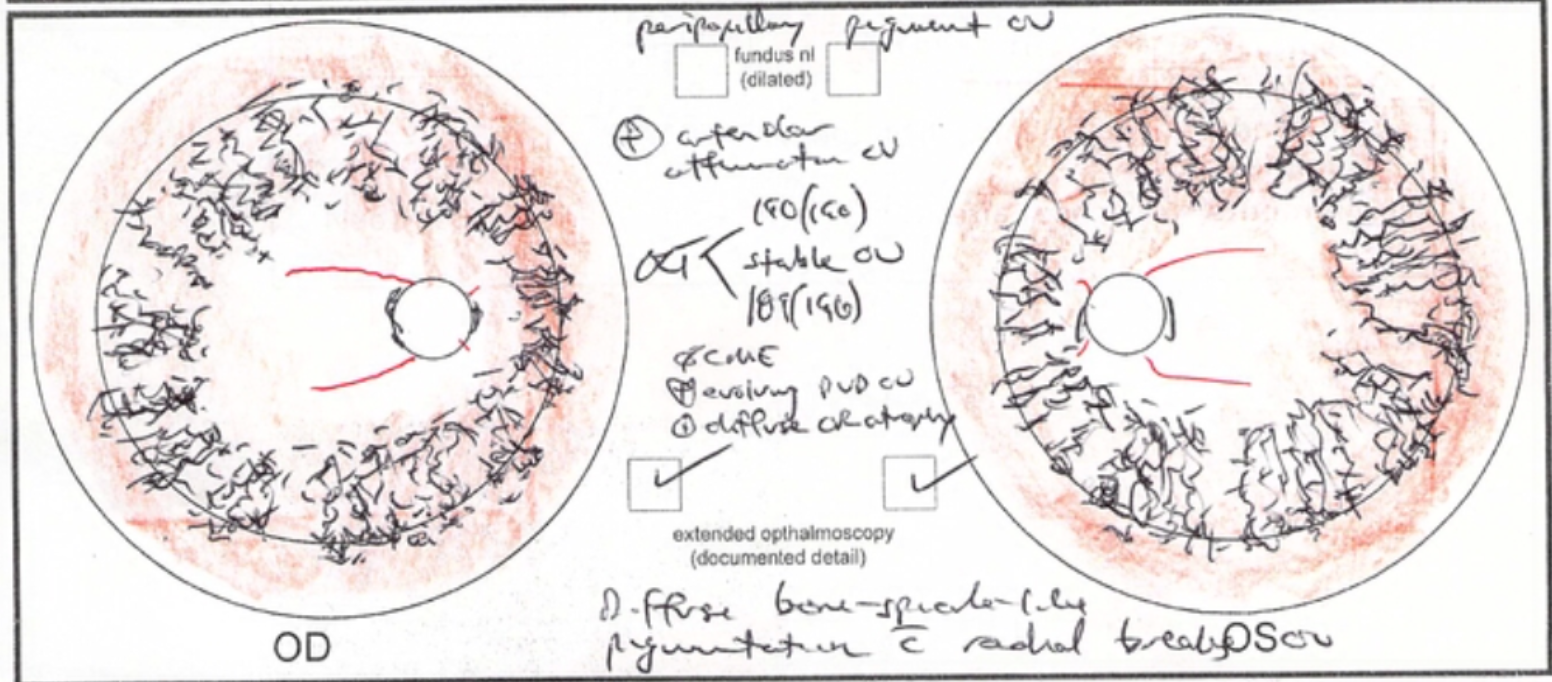
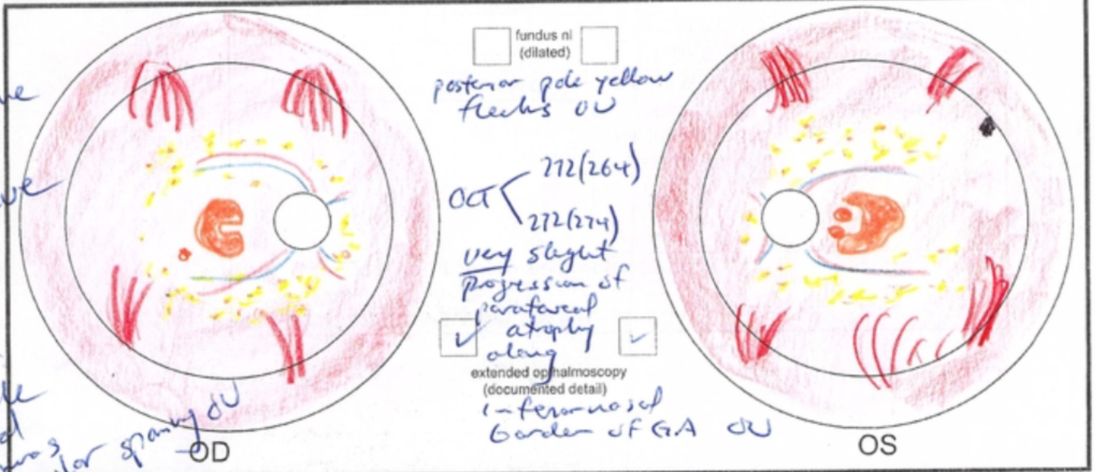
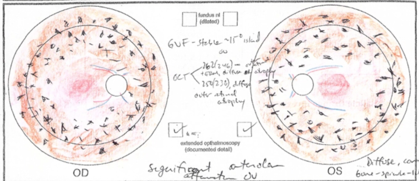
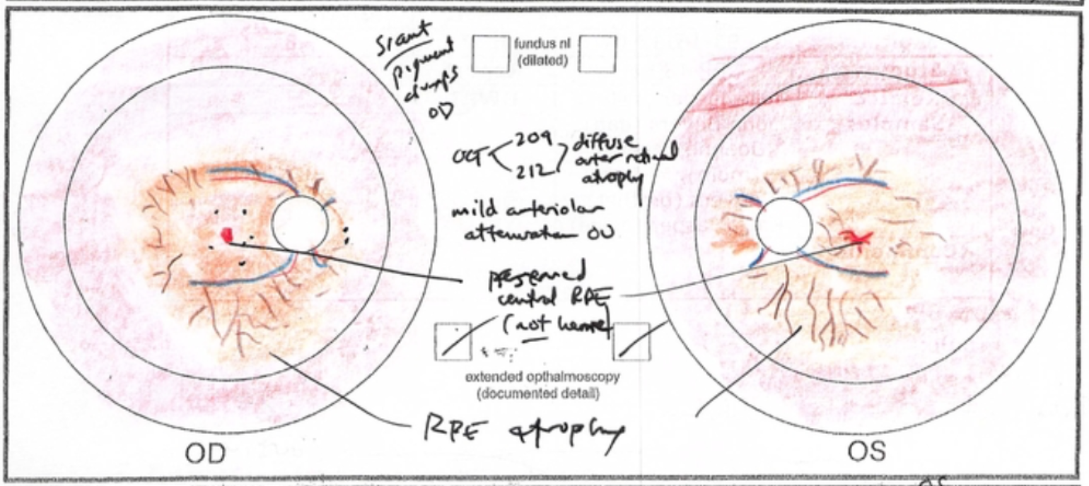
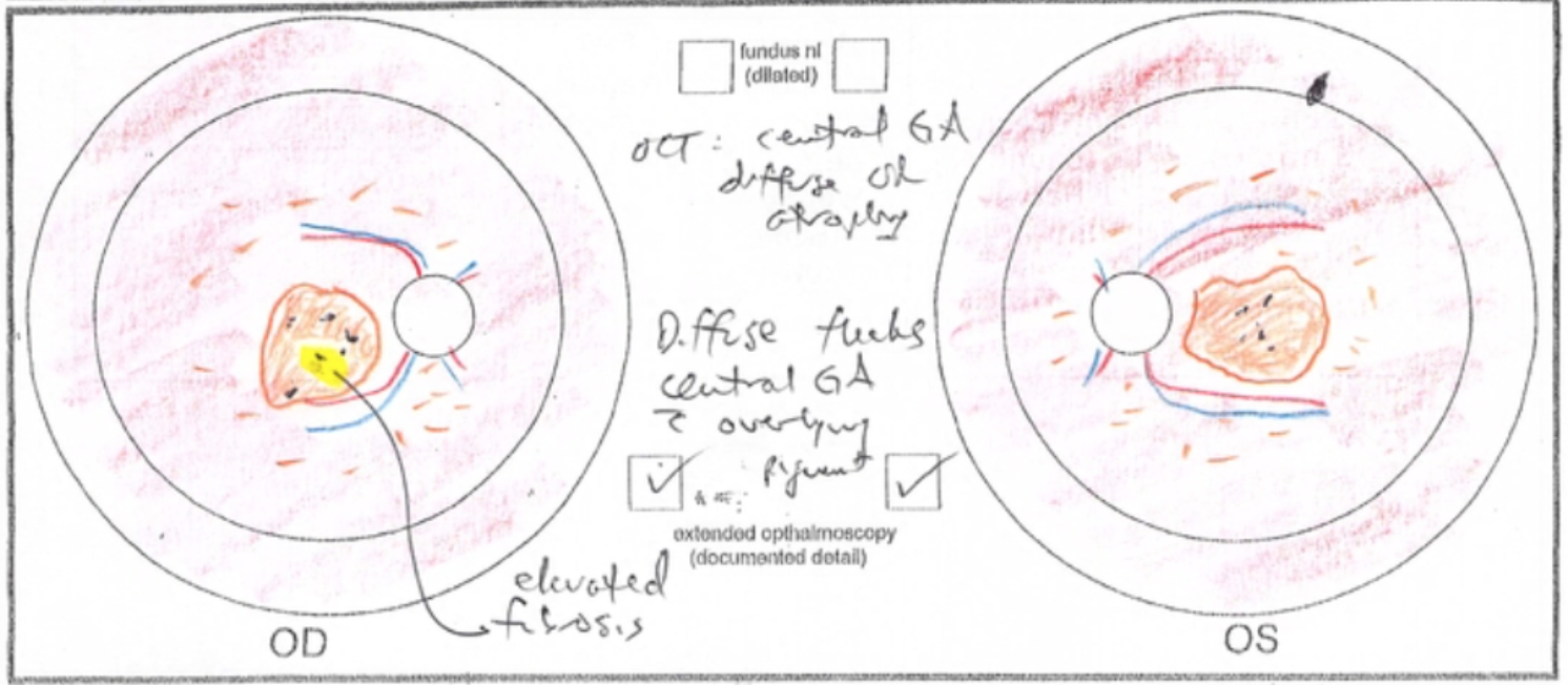
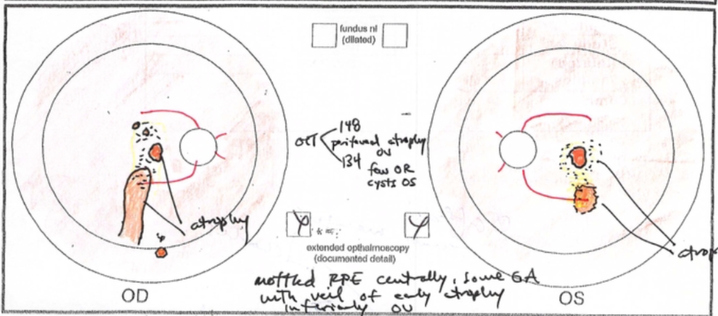
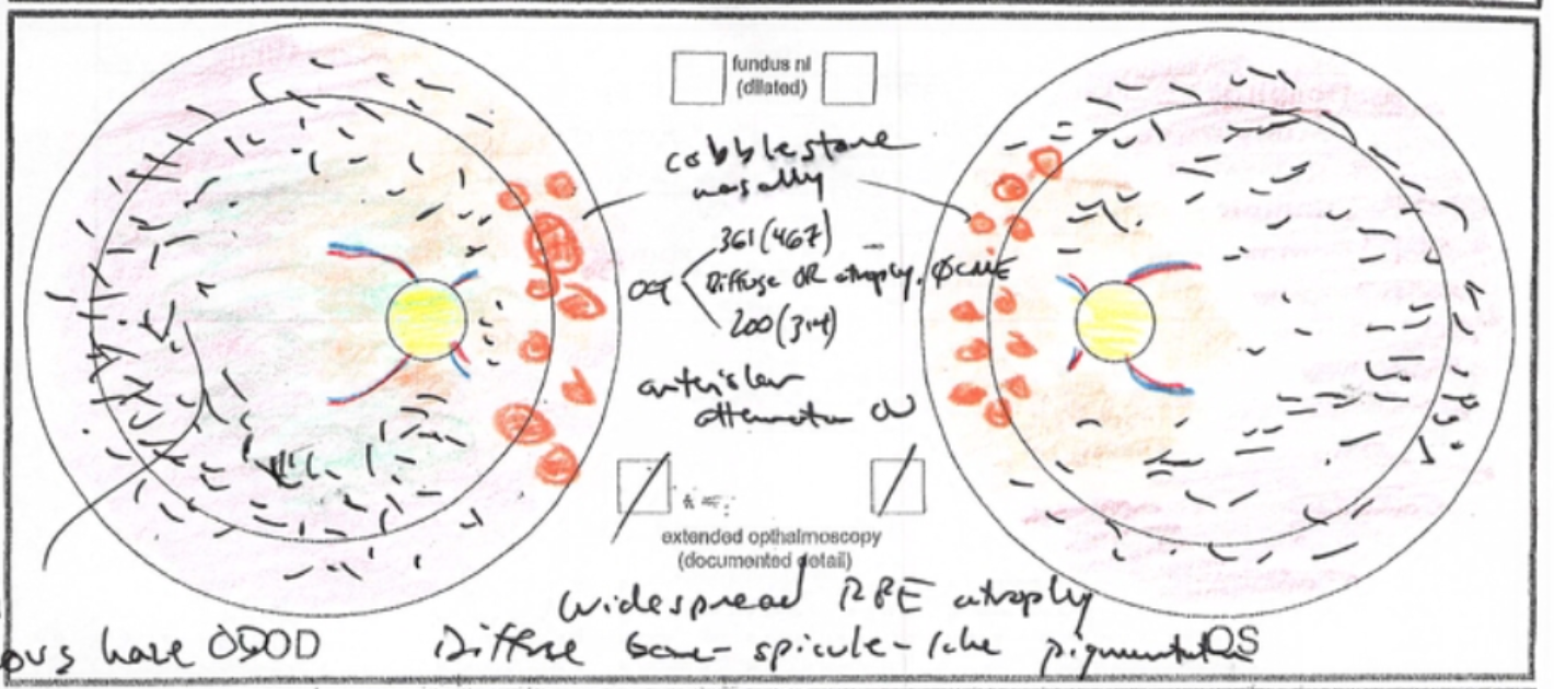
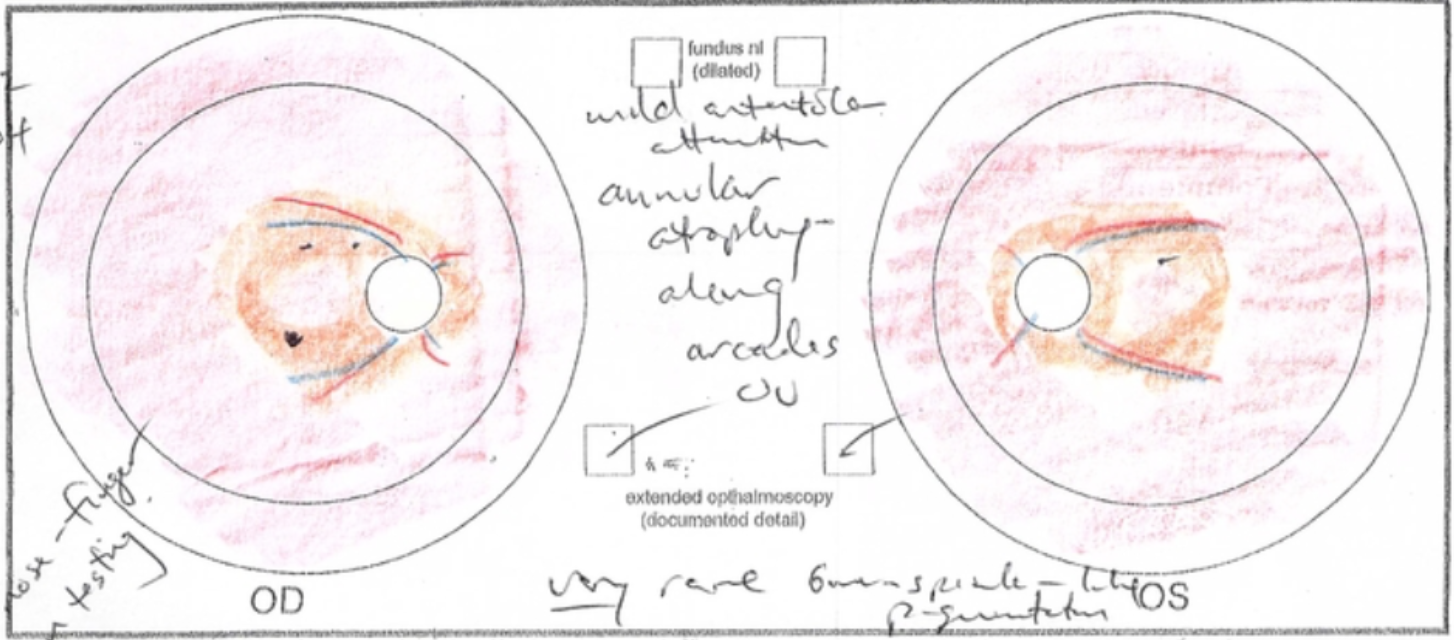
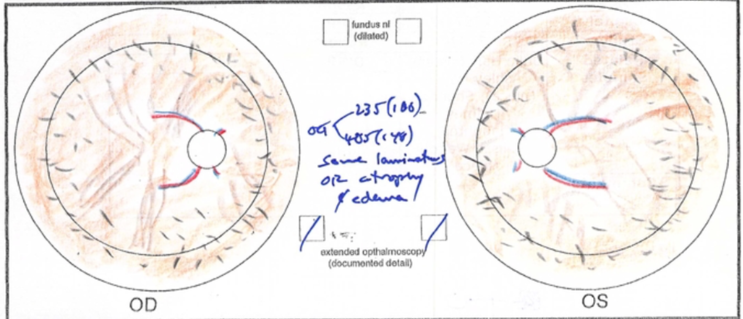
Take a look at my own comparatively very unimpressive retinal art in this slideshow, and then scroll down to see a showstopping example from Dr. Russell's book.